
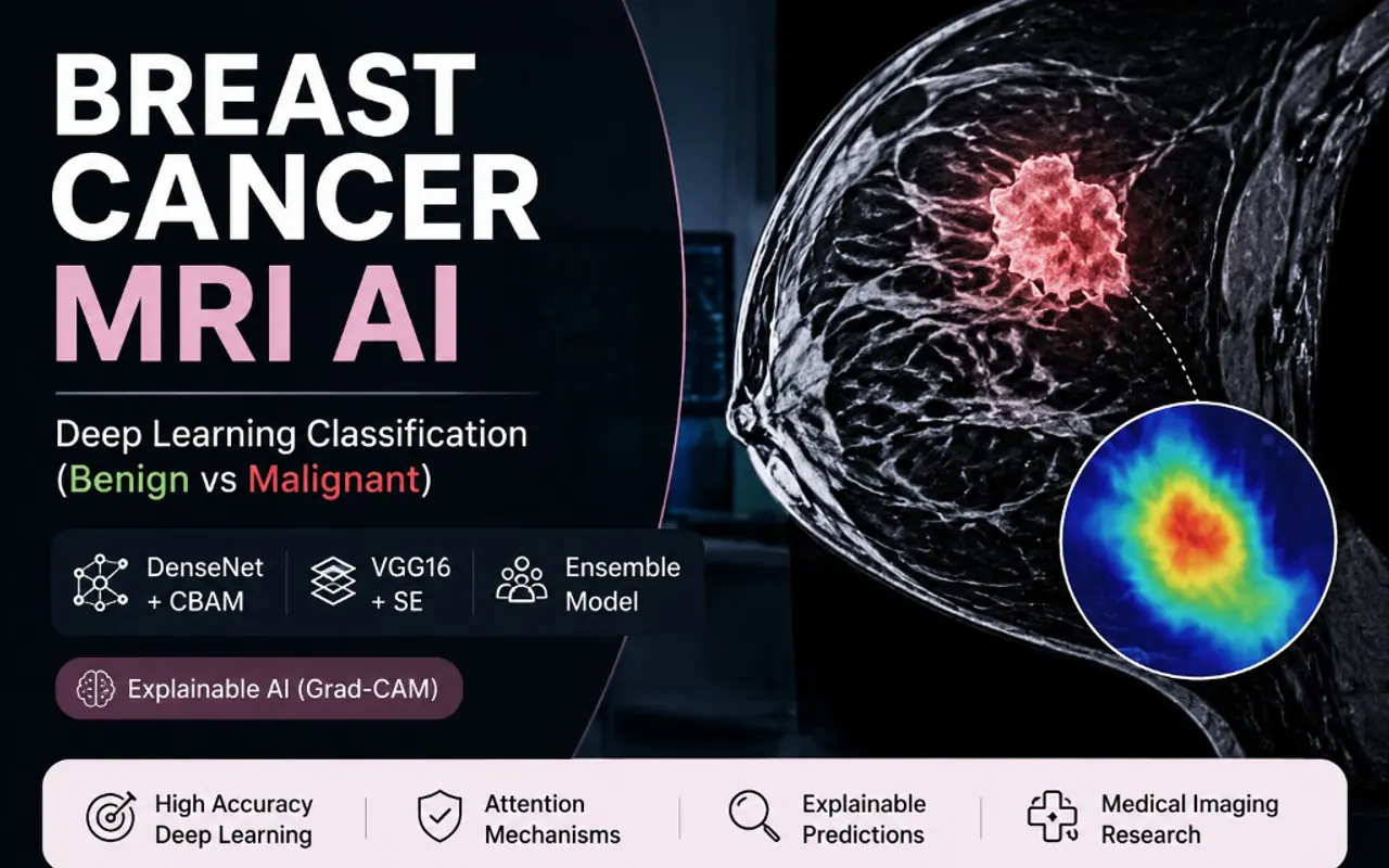
Attention-Based Deep Learning for Breast Cancer MRI Classification
Abstract
A deep learning pipeline that classifies benign vs malignant breast MRI scans using attention-enhanced DenseNet and VGG16, with visual explainability for clinical transparency.
Introduction
Breast cancer is one of the most common and serious cancers affecting women, and catching it early makes a real difference to outcomes. Standard checks like mammograms, ultrasound, and biopsies are the clinical mainstays, but each has limits, some tumours are hard to see, and biopsies are invasive and slow.
Image software can support this work by learning the subtle differences between harmless and cancerous tissue on scans, helping flag concerning cases faster and more consistently. This project builds a system that sorts breast scans into benign or malignant.
Problem Statement
Reading these scans reliably is genuinely hard:
- The differences between harmless and cancerous tissue can be very subtle.
- Results can vary between readers: adding delay and inconsistency.
- The findings need to be trustworthy enough to support clinicians: not replace them.
- A prediction alone is not enough: clinicians need to see why the system reached it.
Objectives
- Build a system that sorts breast scans into benign or malignant.
- Compare a few strong image models and combine them for steadier results.
- Show which regions of a scan drove each decision, for transparency.
- Judge the system on measures that matter in a diagnostic setting.
Dataset
The work is built around a set of breast scans labelled benign or malignant. Depending on the client, this can be a public medical-imaging collection, licensed or private hospital data, scans from other trusted sources, or a custom set gathered for the project. Images are prepared consistently before modelling, and patient privacy is respected throughout.
Methodology
The project trains two attention-enhanced image models, DenseNet+CBAM and VGG16+SE, then combines them in an ensemble so the overall decision is steadier than any single model. Alongside each prediction, Grad-CAM produces a simple visual highlight of the regions that influenced the result.
The novelty is pairing reliable classification with built-in transparency. Rather than a black-box yes/no, the system shows clinicians where it looked, helping them confirm it is focusing on plausible tissue rather than irrelevant artefacts, which builds the trust needed for any real diagnostic aid.