
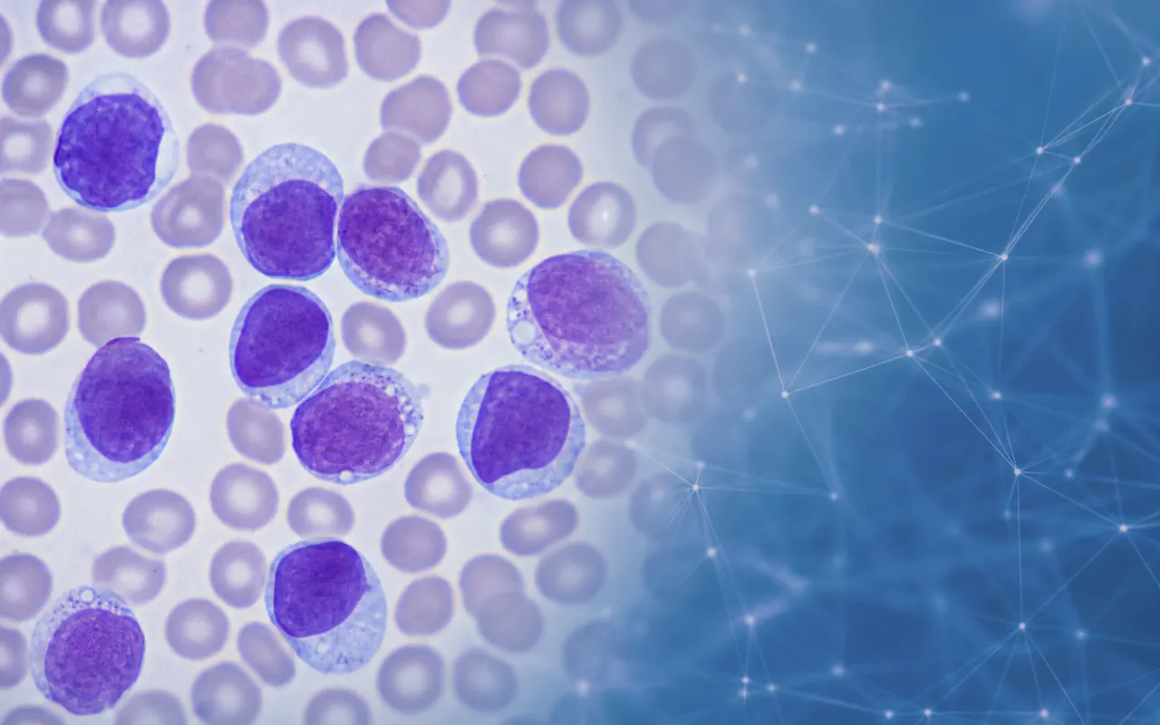
Acute Lymphoblastic Leukemia Classification from Blood Smear Images
Abstract
A deep learning study that classifies benign hematogones versus three ALL lymphoblast subtypes (Early Pre-B, Pre-B, Pro-B) from peripheral blood smear microscopy, comparing Modified AlexNet, VGG19, and DenseNet-121.
Introduction
Acute Lymphoblastic Leukemia (ALL) is a serious blood cancer where early diagnosis depends on examining blood smear slides under a microscope. Doing this by eye is slow, tiring, and easy to get wrong, especially because early cancerous cells can look very similar to healthy ones.
Smart image software can support this work by learning the subtle differences between healthy and cancerous cells, helping labs screen suspected cases faster and more consistently before further confirmation.
Problem Statement
Reading blood smears reliably comes with real challenges:
- The tests are time-consuming: and busy labs quickly build up backlogs.
- Early symptoms are easy to confuse with common illnesses: which can delay diagnosis.
- The different cancer stages look alike: so telling them apart needs expert eyes.
- Mistakes are costly: since a missed or wrong call directly affects a patient.
Objectives
- Build an automated way to screen blood smear images for signs of ALL.
- Tell healthy cells apart from the different cancerous stages.
- Compare a few proven image models to see which works best.
- Keep the approach practical enough to support real lab workflows.
Dataset
The work is built around a dataset that suits the problem well. Depending on the client, this can be a public medical-image collection, licensed or private hospital records, data from other trusted sources, or a custom set gathered for the project. The images are cleaned, balanced, and lightly expanded with standard adjustments before modelling, and patient privacy is respected throughout.
Methodology
The project trains and compares three models on the same prepared images: Modified AlexNet, VGG19, and DenseNet-121. Each learns to recognise the tell-tale look of healthy and cancerous cells, with images prepared and balanced the same way so the comparison stays fair.
What makes the approach valuable is its focus on the hardest part of the problem, separating the early cancer stages that look almost identical, rather than settling for a simple healthy-vs-cancer split. By putting several trusted models side by side under the same conditions, it highlights which one offers the best balance of reliability and practicality for real screening support.